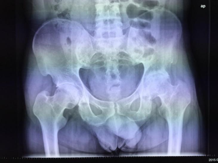
X-ray image before the patient took Collagen Type II, it could be seen that his right side of joint was normal and the space between femoral head and acetabular fossa was also apparent. However, the space between left side of femoral head and acetabular fossa was blurred, which was both jagged and rubbing against each other.

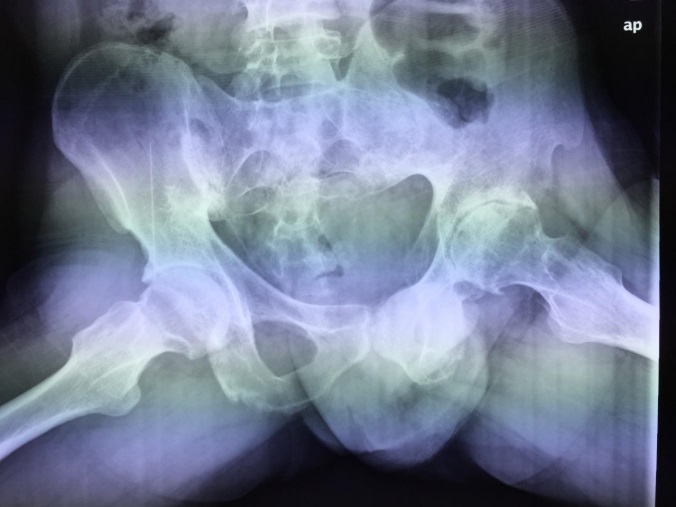
X-ray image after taken Collagen Type II four months, the patient’s left side of femoral head with lesion improved significantly, with some space in his femoral head and acetabular fossa, turning smooth on the surface of bone thus the friction of which diminished.